
Separation of confusion pairs: Eleocharis…
palustris versus austriaca
Shared features
➢ robust and often tall-stemmed plants (though both very variable)
➢ patch-forming or even sheet-forming
➢ spikelets typically many-flowered – at least 20 and often many more flowers/nuts
➢ the two basal glumes are similar in size, short, and typically each encloses just half the spikelet-base with no, or a little, overlap (unlike uniglumis and multicaulis)
➢ stigma 2-forked (as in uniglumis; contrast 3-forked multicaulis)
➢ style-base is enlarged – although to different degrees – and is clearly ‘pinched in’ into a ‘neck’ at the junction with the nut
Separation
These topics (below) are covered. Select a button to go straight down to a topic, or scroll down.
Use the linked "⇑" up-arrows with each topic header to get back here, or use the ‘^’ button which appears in the lower-right corner when you're further down the page.
Links to all pages are at the bottom of the page (wider screens only).
The often-quoted difference in shape of spikelets between these two species is often over-emphasised; it is far from diagnostic!
When nuts are ripe, the glumes become splayed out so that the heads are fatter, and palustris can then have heads as ‘conical’ as austriaca.
Typically, austriaca has conical heads (with +/- straight outlines), or pine-cone-shaped (ovoid-conical); palustris has anything from cigar-shaped, through lanceolate, to ovoid and ovoid-conical.
Don’t despair! - read on...
austriaca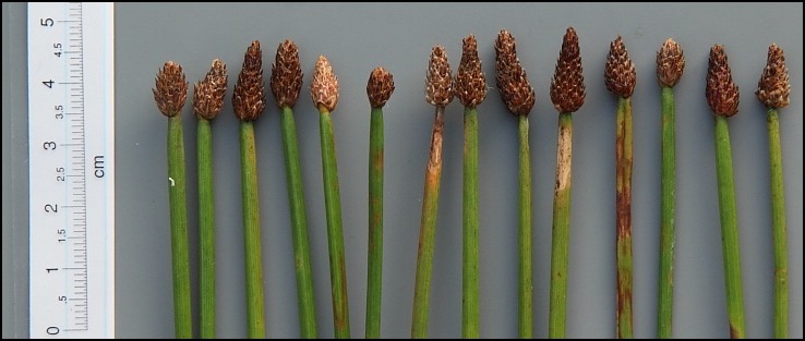
palustris (typical, early-season, with cigar-shaped and lanceolate heads)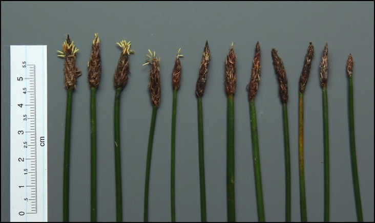
palustris (late season, a form with short, ovate heads closely matching some austriaca)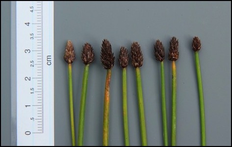
Style-base (stylopodium) ⇑
The most immediate and readily-discerned character for field separation of this pair once nuts begin to develop.
The style-base in palustris is invariably more swollen and bulky than that in austriaca, although there is variation in each.
austriaca NUTS palustris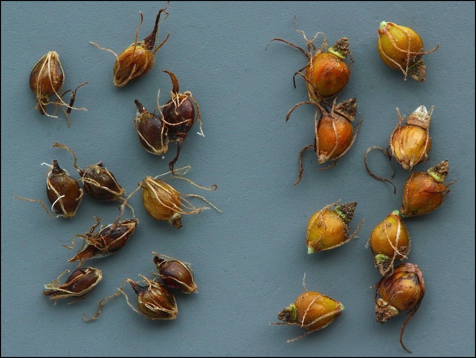
Perianth bristles ⇑
palustris | austriaca | |
No. perianth bristles | 4 (almost invariably) | 5 (rarely 4 or 6) |
The number of bristles is a good distinction, and useable over a long period of the summer and autumn as the bristles are persistent at the base of the nut.
(On an inspection of the base of the nut with a 20x lens, the bristles are fairly easy to see arising from a whitish collar at the base of the nut. A count of bristles on five nuts should be sufficient.)
Bristles in austriaca generally appear longer than those in palustris. This may well be a good distinguishing feature, but the bristles are somewhat brittle, and being flexuous it can be difficult to judge the length - and certainly impossible to measure conveniently.
Beware! Three +/- persistent stamen-filaments also arise from the base of the nut, but emerge from inside the perianth-collar, and so adjacent to the nut. These tend to become darker brown with age, whereas the bristles remain paler brown. Filaments are rather broader than bristles and lack the minute down-curved hooks distinctive of bristles. (The minute hooks act to cause the nuts to cling to each other, and to animal or bird vectors, aiding dispersal.)
The photographs of nut-bases below show (i) the four bristles of palustris clearly (but the filaments have been shed), but (ii) the austriaca picture (filaments still present) is less clear and needs some labels.
palustris (4 bristles, 0 filaments ~ shed)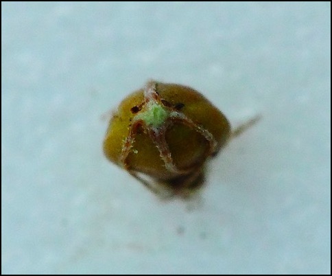
austriaca (5 bristles, 3 filaments) 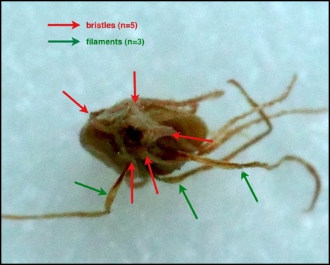
The following palustris diagram shows all the features: the swollen style-base with ‘neck’, four bristles and three filaments. The style-plus-stigma has already been broken off. The whole is about 2 mm long.
palustris 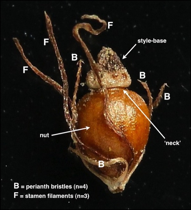
The following austriaca diagram shows two adaxial (outer-side) bristles arising directly from the basal collar....
austriaca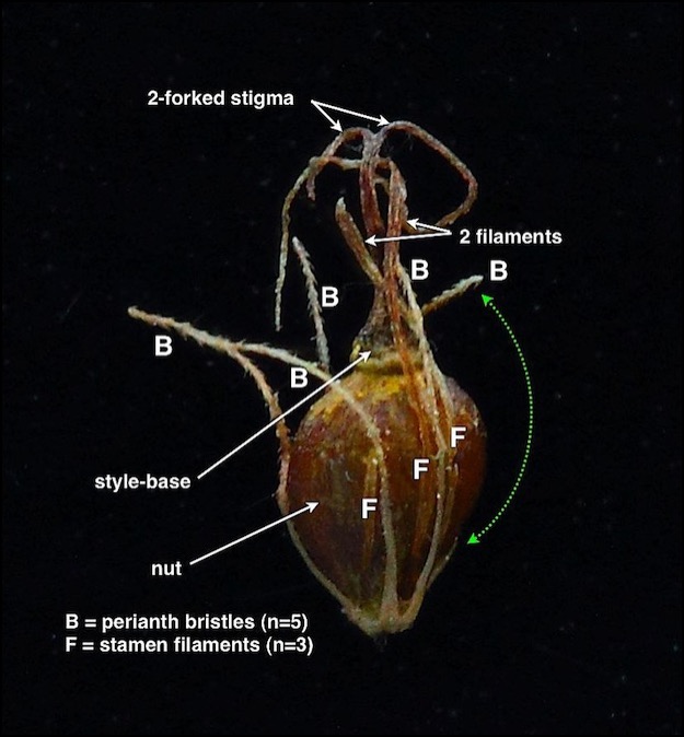
....whilst the next shows a single adaxial bristle forked near the base. Such a forking bristle is counted as two bristles.
austriaca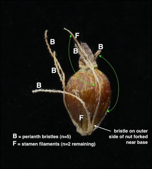
Thus in austriaca the count of five bristles arises through forking of the bristle on the outer side of the nut, which may occur at the very base of the bristle (thus generating five clearly distinct bristles), or at some higher point (making a +/- Y-shaped outer bristle). To reiterate, this forked bristle is still counted as two.
Occasionally, six bristles arise: the following picture shows six bristles (two from a confluent base) and three filaments in austriaca - nine structures in total!
austriaca (with 6 bristles)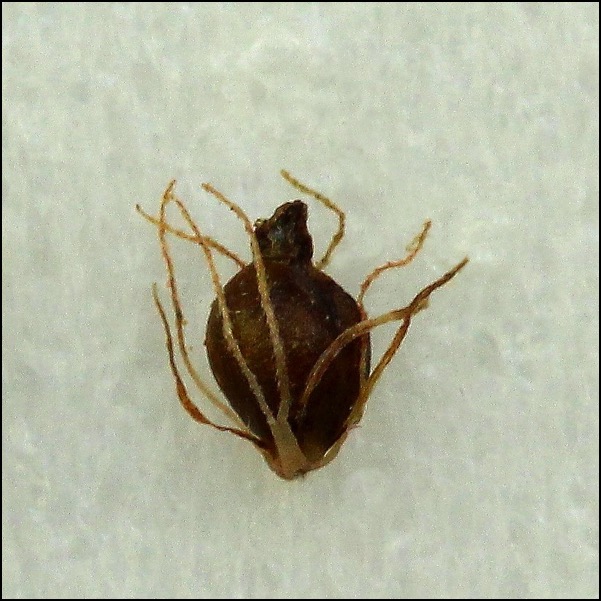
Stem ⇑
Characters are variable: palustris can match austriaca at times:
| STEM… | palustris | austriaca |
| … section | often slightly flattened or oval | circular |
| … colour | often dark green (but can be pale) | often bright- or yellowish green |
| … colour at base | often flushed red/purple | usually pale green |
Stem section ⇑
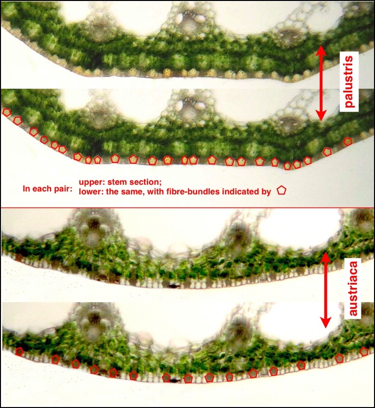
In section, and at the same scale, palustris can be seen to have deeper tissue layers below the epidermis (outer skin) – almost twice the thickness – and the oval section is obvious, versus the almost circular section typical of austriaca.
austriaca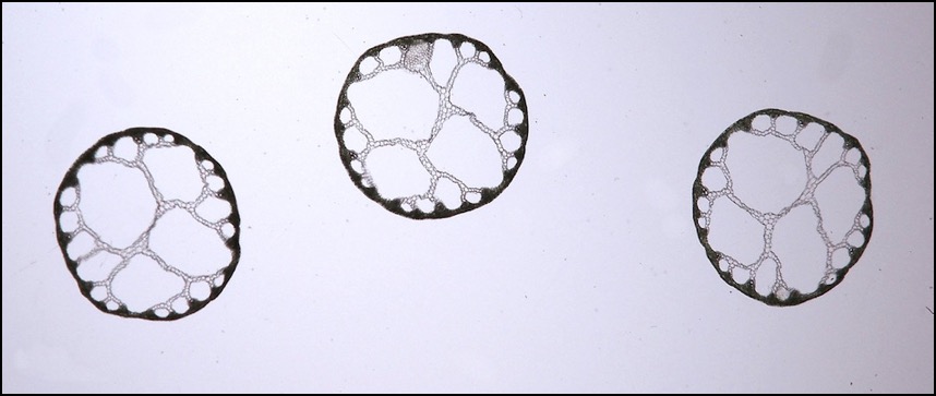
palustris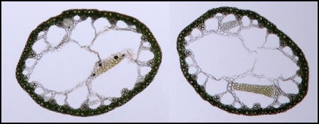
In a closer view (below) of small parts of the stem sections, the outer layer – the epidermis – can be seen (just about) to be made up of alternating bundles of fibre-cells, about pentagonal in section, and rows of clear cells. In austriaca the bundles of fibre-cells are more regularly spaced and separated by wider strips of clear cells made up of more rows than in palustris.
The fibre-bundles are indicated in red in the lower of each of the pairs of photographs (palustris: upper two; austriaca, lower two).
These differences can be visualised more readily if an epidermal peel is prepared - see next.
Epidermal peel ⇑
With a microscope, one of the best distinctions is the fine structure of the epidermis (the outer layer) of the stem. Hence purely vegetative material of putative austriaca may be identified with full confidence: no more excuses for ignoring non-flowering plants!
This can be revealed by a process which needs some practice to produce a good ‘peel’. (Dried material will need a section of stem boiling for about 5 minutes to soften it.)
Try this (and let me know if you find a more reliable technique!):
1) Slice a section of about 5 cm of moist stem longitudinally to produce two ‘gutter-shaped’ halves.
2) Lay one slice on a glass slide, outer surface downwards, and add a drop of water.
3) Hold the piece firmly down with a forefinger, and with the other hand place a razor-blade down vertically across the slice touching the finger-end.
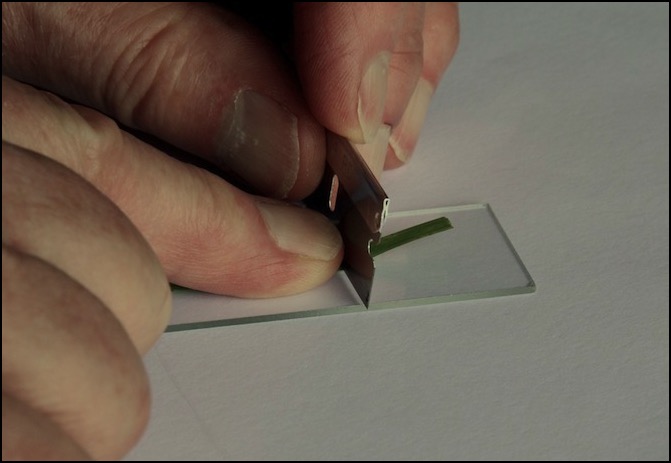
4 i) With luck, a series of careful scrapes along the strip may remove green tissue and leave colourless epidermis. Make several ‘passes’ and scrape off more green tissue with each pass. The process is easier under a dissecting microscope.
4 ii) Sometimes it needs more care. Whilst holding the blade vertically, the sharp edge exactly parallel to the slide, and at ‘just the right height’ (only determinable through experiment) push the blade along the strip, scraping away green tissue and leaving transparent epidermis.
This is most easy to achieve by a steady forward push with the retaining finger-end, using the flesh of the finger both to keep the slice in place and to push the (vertical) blade very slowly and firmly along the slice, pushing green tissue in front of it and leaving transparent epidermis behind. Only one or two square millimetres of clear epidermis may be enough to elucidate the features.
Blade height is critical:
~ blade too low: tearing of the epidermis is likely
~ blade too high: some masking green tissues remain
~ blade angled forward: slicing into the epidermis is likely
~ blade angled backwards: green tissue left behind
~ blade edge not parallel to slide: thinner edge likely to tear away
5) Invert the peel to bring the outer surface uppermost, add more water and a coverslip.
You should then be able to see the features as shown next.
These photos are at the same scale. The length of stem in this view (i.e. across the screen) is about 0.8 mm.
The key on the right of each photograph shows:
- RED lines indicating the position of bundles of elongated fibre-cells within the epidermis. These contribute to the strength and pliability of the stem.
- GREEN figures indicating the number of rows of ordinary thin-walled epidermal cells between bundles of fibre-cells. The stomata (the breathing pores: little ‘mouth-like’ structures) are situated within these rows.
It is clear that palustris has far more fibre-bundles (in places so dense as to be difficult to discern how many bundles!), and narrower strips of ordinary cells, mostly only 1-3 cells wide....
palustris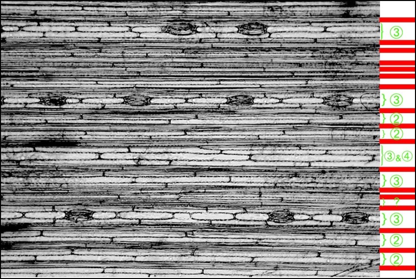
.... By contrast austriaca has fibre-bundles fewer, never bunched, and much more regularly spaced, with bundles separated by strips of ordinary cells 3-5 rows wide.
austriaca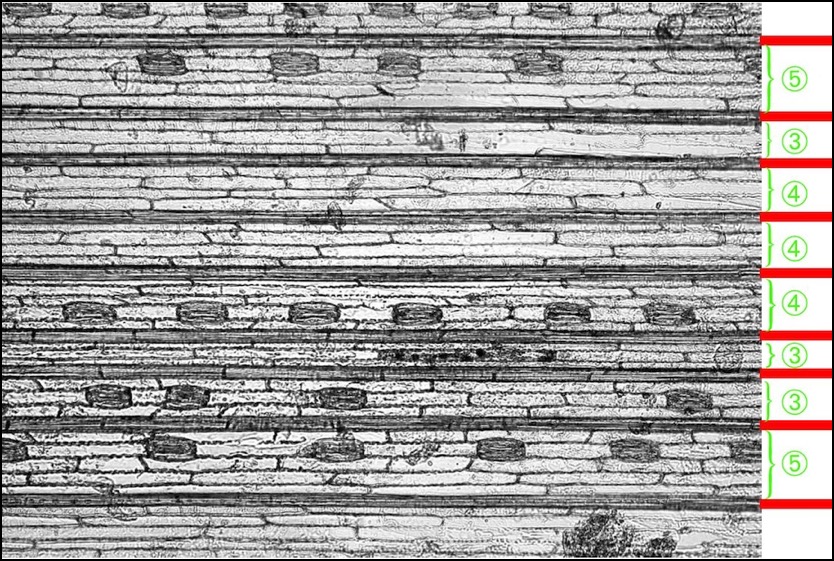
Stomata ⇑
The length and shape of the stomata are excellent diagnostic characters to tell austriaca from the common UK plant palustris (subspecies vulgaris).
(The rare, southern, Eleocharis palustris subspecies palustris has shorter stomata than in subspecies vulgaris, and the shape is also closer to austriaca - at least to judge by the photos in Strandhede (1966). However, as far as we know, there is no overlap in the UK between the northern range of austriaca and the southern range of subspecies palustris. Of course, many other characters will separate these two.)
Stomatal length
Although there is much overlap in length, in practice (because these are mean figures) many specimens can be separated on this character (figures from Strandhede & Dahlgren (1968)):
➢ in austriaca the mean range is (38–) 42–52 (–65) μm
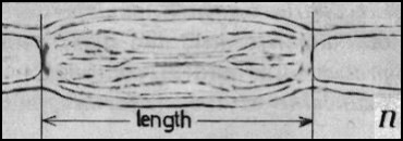
Strandhede (1966) provides a diagram illustrating the precise measurement taken (right).
The critical difference in shape is in the lengths of the 'guard' cells (the pair of cells forming the stoma, the opening, itself) versus the lengths of the 'subsidiary' cells (the pair of cells lying alongside the guard cells)
➢ in palustris subspecies vulgaris, guard cells are shorter than subsidiary cells
➢ in austriaca, guard cells are longer than subsidiary cells
This can be seen in the photos below.
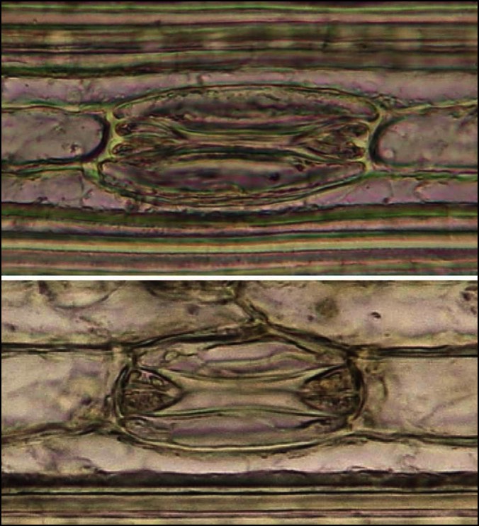
(upper) palustris subspecies vulgaris
(lower) austriaca
Whilst the actual comparative lengths of the cells (guard vs. subsidiary) can often be difficult to judge (these photos are my best efforts at illustration so far!), the differences are more obvious from their impact on the adjacent cells within the same row at each end of the stomata. Thus:
- in palustris (upper) these adjacent cells have convex ends to the lumens because stomatal cells are shorter than subsidiary cells
- in austriaca (lower), stomatal cells are longer than subsidiary cells and hence 'bulge' into these lumens.
See the key, next.
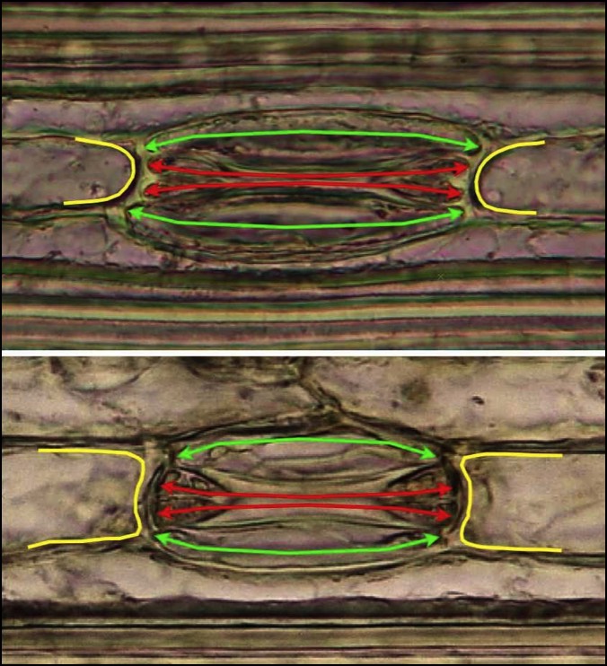
~ the lumens of the guard cells arrowed in RED
~ the lumens of the subsidiary cells arrowed in GREEN
~ the margins of the lumens of the adjoining cells at each end of the stomata in YELLOW
(upper) palustris subspecies vulgaris
(lower) austriaca
In each, note comparative lengths of green subsidiary cells vs. red guard cells.
Links to the other Eleocharis spike-rush pages (also accessible from the sidebar)
Species pages
Separation of similar pairs
Other information